Nine years is a long time in tomography.
In 2017/18 the first UKRI UK X-ray Tomography Roadmap defined the nations landscape of hardware and software, directly driving the creation of the UKRI National Facility in X-ray CT (NXCT) to broaden access to national infrastructure.Today, the landscape has transformed. Facilities have evolved and more established nationwide, the user base has grown and broadened, and has resulted in growing demands for skills. With these developments, the roadmap requires a refresh. It shifts the focus from equipment ownership to access, hardware locations and software distributions will be remapped, considers skills, and will culminate in defining strategic priorities for the future.
- Last roadmap
- 2017/18
- Working Group
- 12 members
- Publishes
- Spring 2027
Why now
The original UKRI roadmap suggested a 3 to 5-year refresh, but without a concrete plan to execute it. Since then, the National Facility opened its doors in November 2020 (!), completed its initial 5-year grant, and has now been renewed as "NXCT II" for another 5 years. Similarly the Collaborative Computational Project on Imaging (CCPi) was refreshed by UKRI with a strong focus on open-source tomography software.As part of their current grant which ends 2027, CCPi agreed to re-run the roadmap exercise to ensure objectives remain aligned with community needs. While 10 years would have been a round number, there is a clear timing alignment across these national initiatives to reassess requirements now.
Who is behind it
The roadmap is chaired by Dr Jay Warnett (University of Warwick) who is Co-I on both the NXCT and CCPi grants. Working group members were nominated following discussion with NXCT and CCPi executive teams with careful consideration given to balancing disciplines, facility sizes, career stages, EDI, and geographical location. Relationships to National centres and initiatives relevant to X-ray Tomography were also considered.This group is responsible for interpreting the data provided by the community via the survey and listening to your views from the town hall to generate a non-biased presentation of national infrastructure capability and requirements. Together, we aim to ensure the UK X-ray tomography community is accurately represented to key funders and policy stakeholders.
Your data, your roadmap
We will be collecting data to inform the writing of the roadmap via two methods. First on online survey is distributed to collect base data from X-ray Tomography users regarding hardware/software access and use, skills development and current challenges facing their work. At the town hall meetings we present the initial findings of this survey. It then provides an opportunity to discuss as a community what are the most pressing needs and challenges, and what we need to overcome them.The working group will use this information to write the roadmap, presenting the current infrastructure landscape and relaying the future requirements and opportunities for X-ray Tomography. This is expected to be published in Spring 2027 in a variety of mediums with citable DOIs.
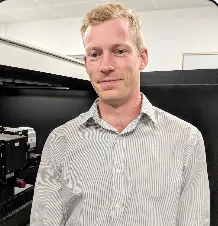
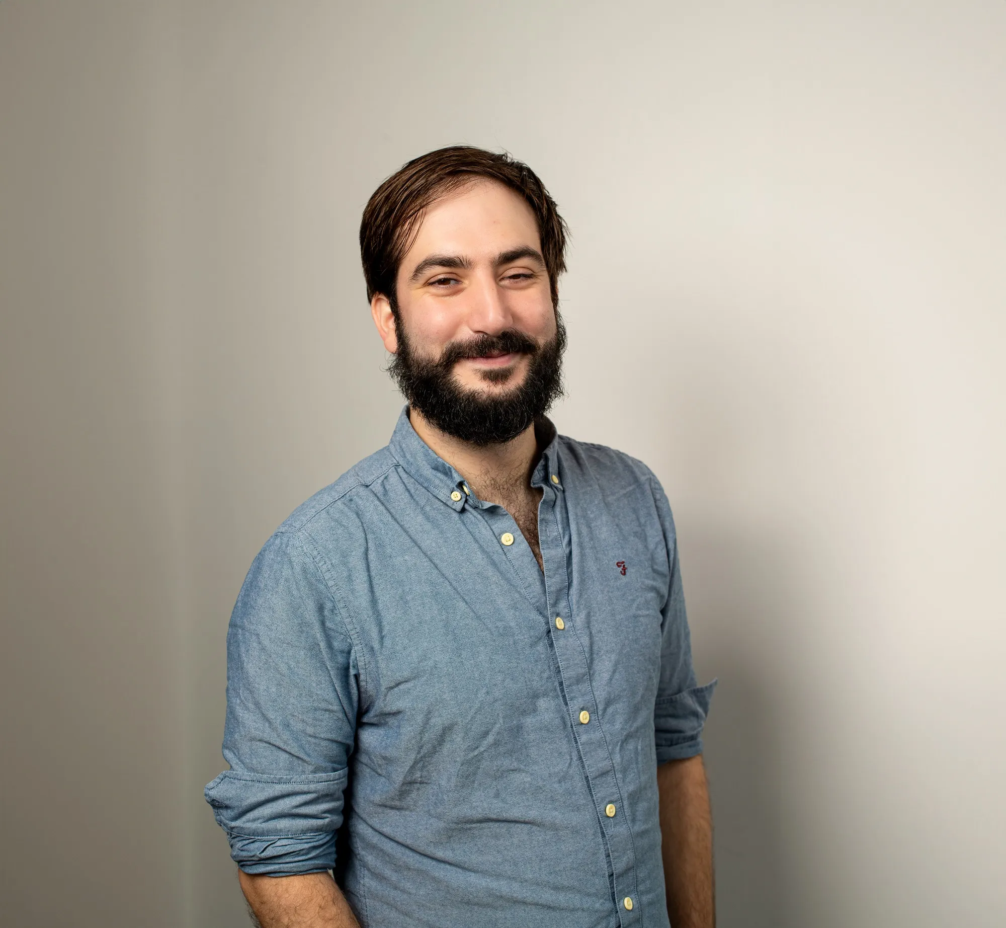
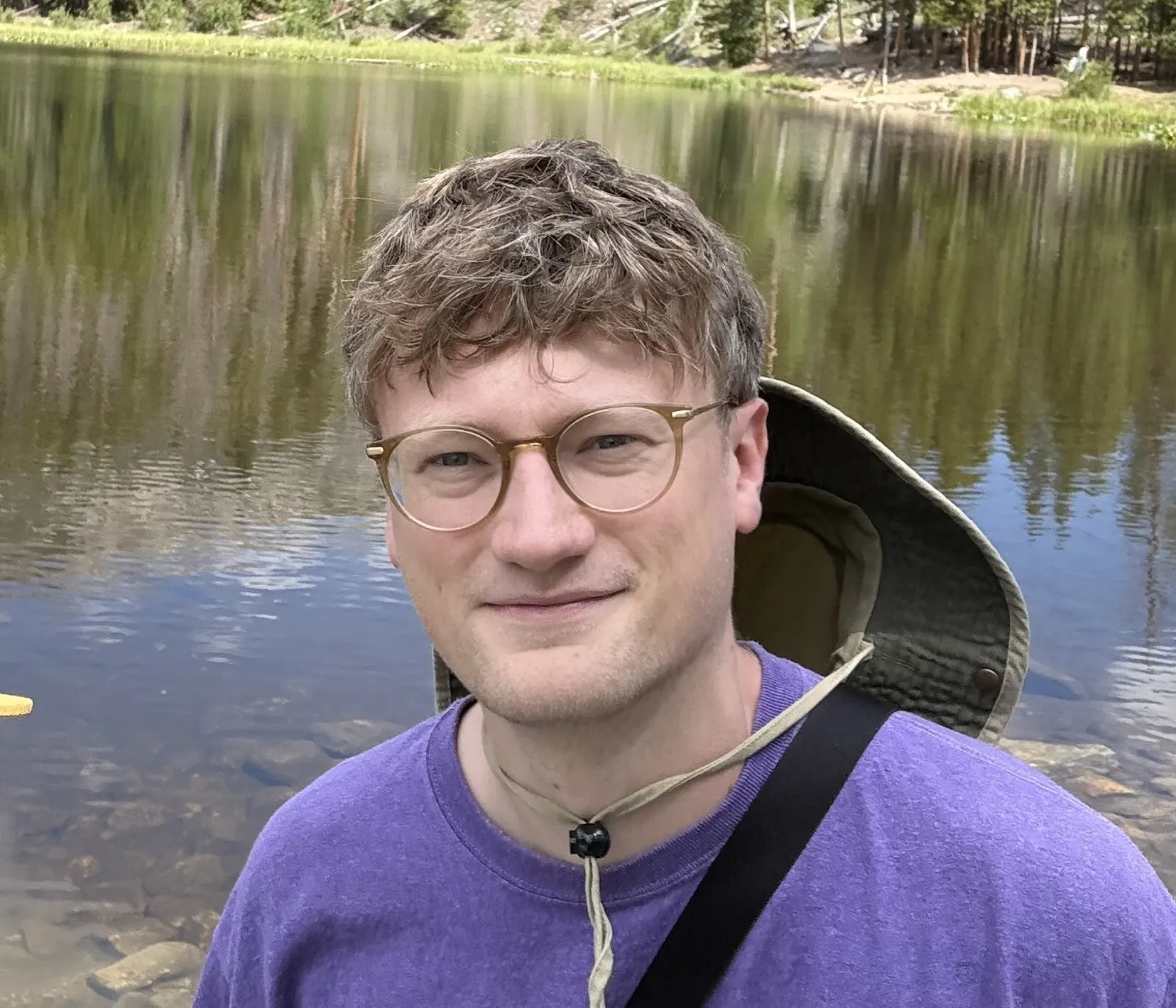
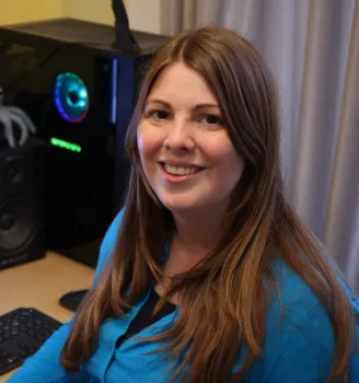
Meet the Team
Contact any of us directly, or write to the group via the chair at [email protected].





What happens to what you tell us
People are candid about their instruments and their frustrations only if they trust where it lands. So, plainly:
Held off-site
Responses go to the survey platform, not to this website. This site is a set of static pages with no database and no analytics on your answers.
Stripped before analysis
Names, emails and free-text that could identify a person or a single facility are removed before anyone in the Working Group reads the data.
Aggregate only
Findings are published as counts and themes. No result is reported where a small group could be inferred, and no institution is ranked or named.
Contact details you give for town halls or updates are kept separately from survey responses and are never joined to them. Ask us to delete either at any time — [email protected].
One technique, hundreds of questions
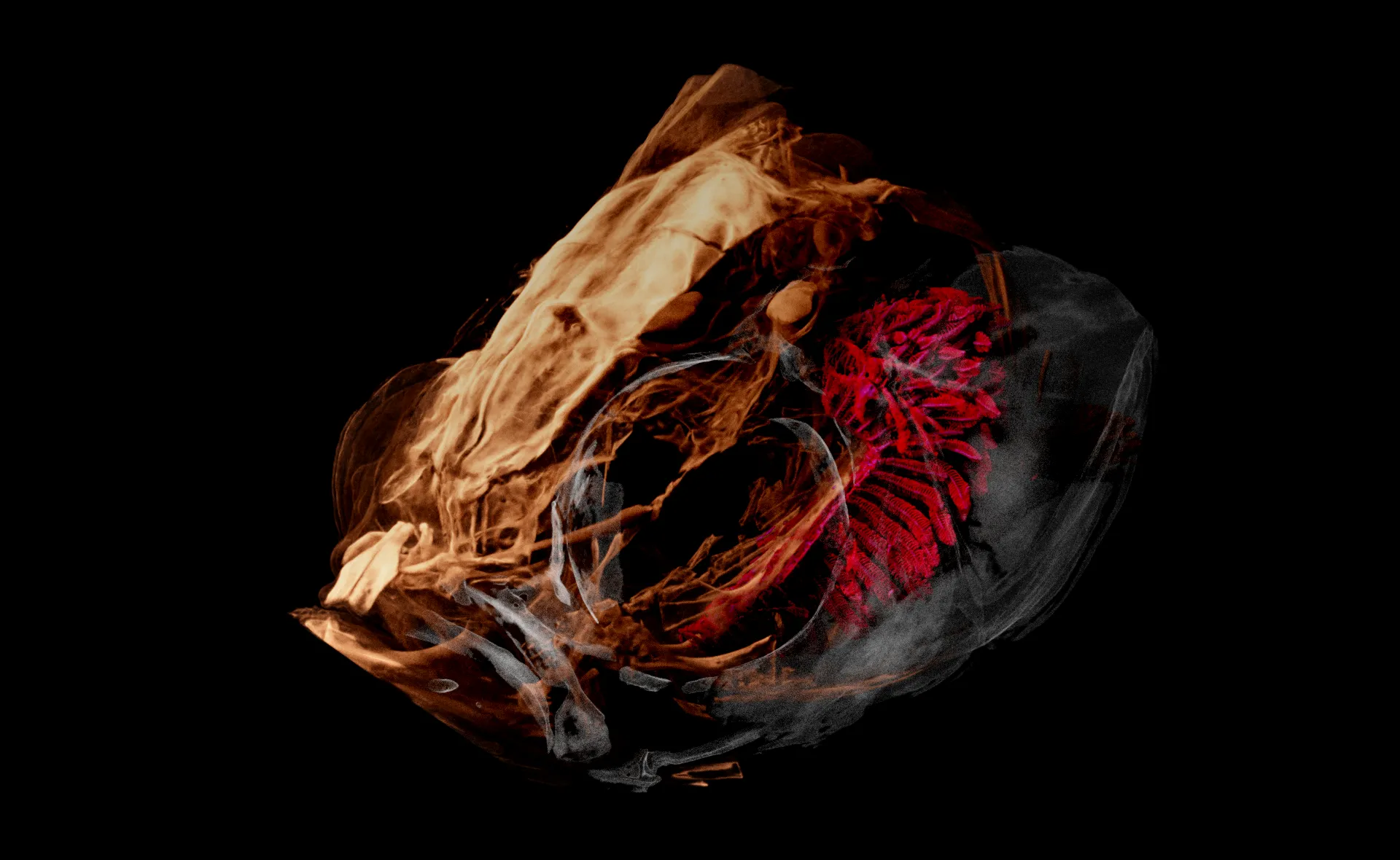
Zebrafish head. Soft tissue and gill structure resolved at micron scale.
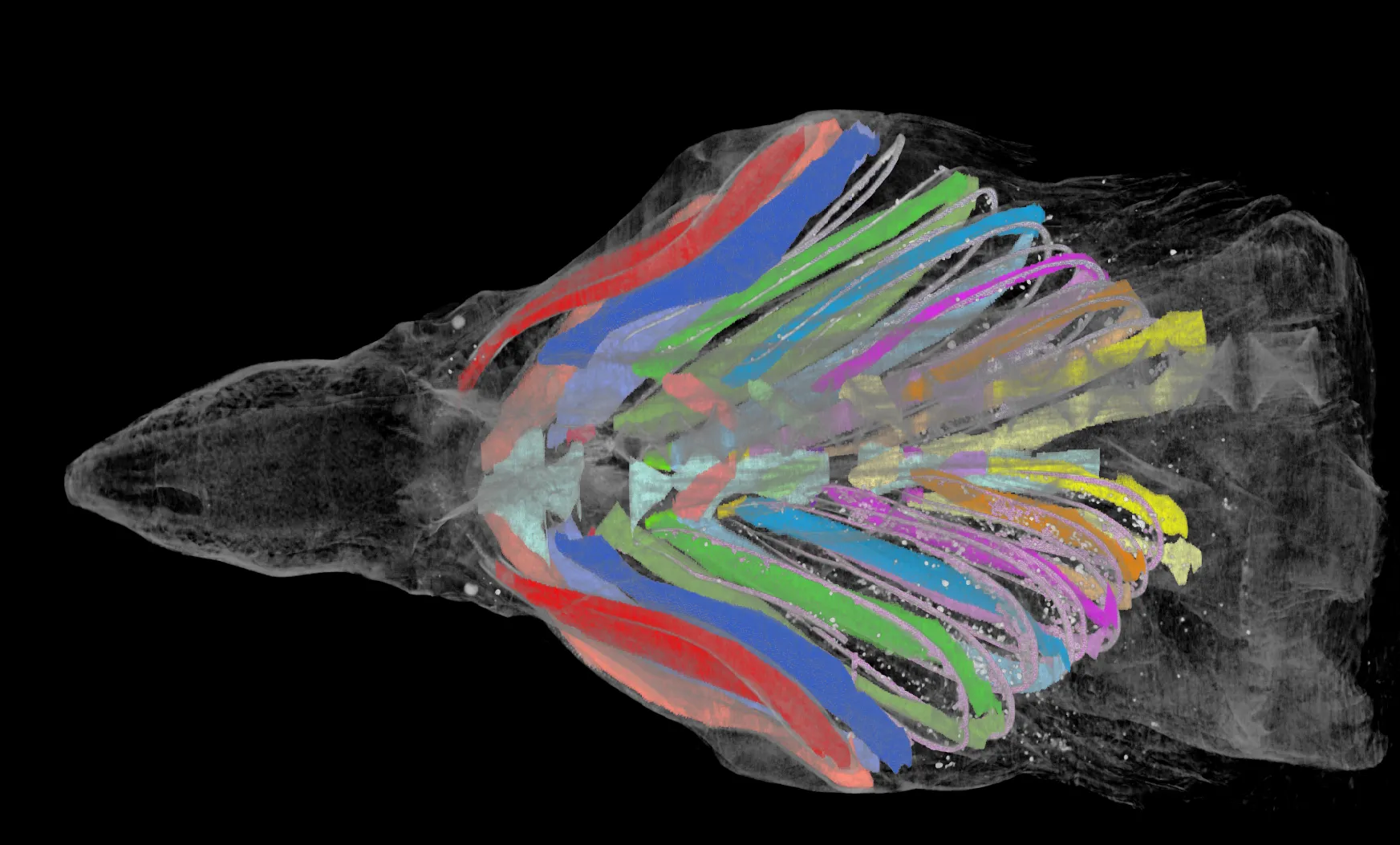
Segmented musculature, laboratory micro-CT, labelled block by block.
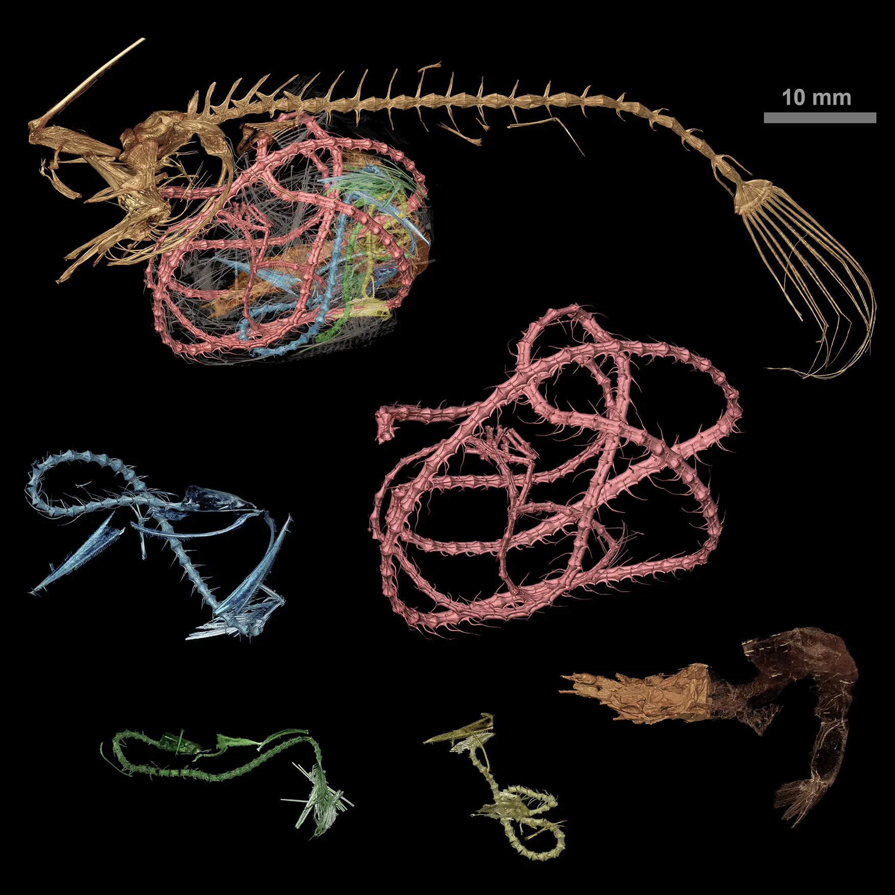
Skeletal elements isolated from a single volume and re-rendered.